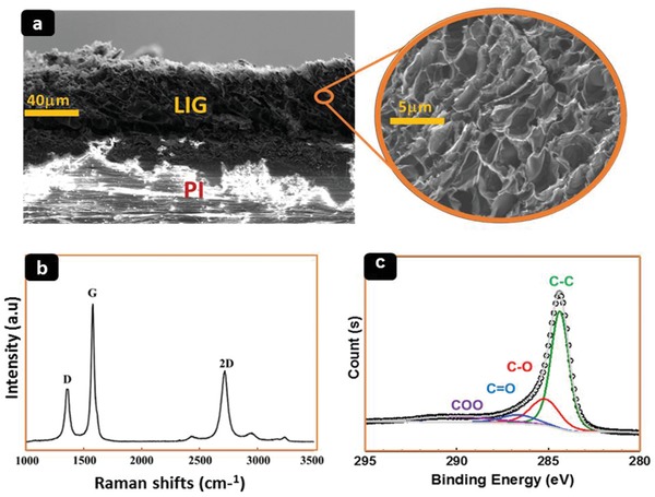
Figure 2.

a) Cross‐sectional SEM images of porous graphene structures on PI after laser irradiation. The inset with a higher magnification shows randomly arranged and interconnected graphene flakes. b) Raman spectrum of LIG acquired with a laser wavelength of 473 nm. c) High‐resolution XPS spectrum of the C1s region of LIG.
