
Figure 5.
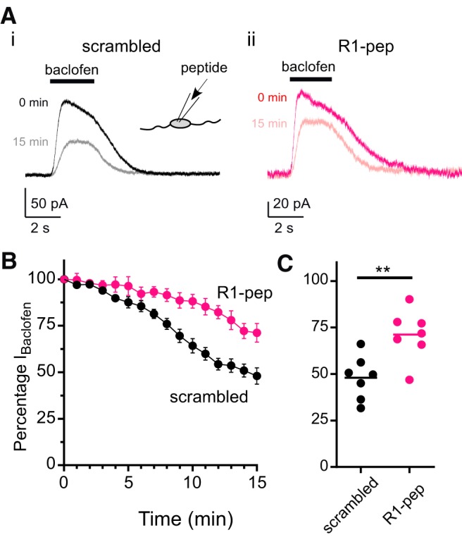
R1-pep peptide attenuates rundown of baclofen-activated GIRK currents (IBaclofen) in cultured cortical neurons. A, Representative traces showing baclofen-activated currents recorded from rat cortical neurons at baseline (0 min) and after 15 min for the scrambled peptide (Ai) (QPRTPRHLSQRR, black traces) and PP2A-interfering peptide (Aii) (R1-pep: RQQLRSRRHPPT, magenta traces). Black bars above traces indicate the duration of the baclofen (10 μm) pulse. Holding potential was −50 mV. Inset, Diagram shows inclusion of peptides in the patch pipette and thus directly exposed to the cell interior. B, Plot of the normalized IBaclofen over time for recordings with control peptide (scrambled, black) and R1-pep peptide (magenta). Mean ± SEM is shown (n = 7 cells per condition). C, Dot plot showing the current remaining at 15 min expressed as a percentage of the baclofen-induced current at 0 min for cells exposed to either R1-pep (magenta) or scrambled peptide (black). Bar indicates mean (**p = 0.0046; t = 3.468, df = 12, two-tailed unpaired t test).