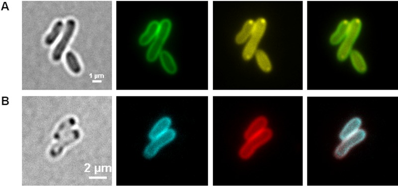
FIG 2.
(A and B) Staining of A. tumefaciens with FM1-43 and DAPI after 24 h (A) and with FM4-64 and LysoSensor green DND-189 after 48 h (B). (A) Cells were grown in LB medium in the presence of 0.1 μg/ml FM1-43. Samples were taken at 0, 2, 4, 6, 24, 32, 48, 52, and 72 h after inoculation, stained with 0.5 μg/ml DAPI, and (from left to right) imaged with bright-field microscopy or with fluorescence microscopy with an FM1-43-specific or DAPI-polyP-specific filter, with the rightmost being an overlay of the green and yellow channels. The same result was obtained if FM4-64 was used instead of FM1-43 together with an FM4-64-specific filter set (not shown). (B) A. tumefaciens wild-type cells were stained after 48 h with 1 μM LysoSensor green DND-189 and 0.1 μg/ml FM4-64 and analyzed with an FM4-64-specific or a LysoSensor green-specific filter. The experiment was performed in three biological replicates. A typical result is shown.

