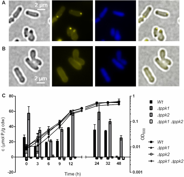
FIG 6.
PPK1 is responsible for polyP formation in A. tumefaciens. (A and B) LB-grown A. tumefaciens Δppk2 mutant cells (A) still formed DAPI-polyP foci, but DAPI-stained A. tumefaciens Δppk1 mutant cells (B) showed no characteristic DAPI-polyP fluorescence signal. From left to right are bright-field, DAPI-polyP channel, DAPI-DNA channel, and merged images. Microscopy images were taken from three biological replicates. (C) Growth on LB medium (OD600) and polyP content in μmol Pi per cell dry weight (cdw) of A. tumefaciens wild type and Δppk1, Δppk2, and Δppk1 Δppk2 mutants. The growth experiment was performed in a plate reader with five technical replicates and the polyP quantification with three technical replicates.

