FIG 8.
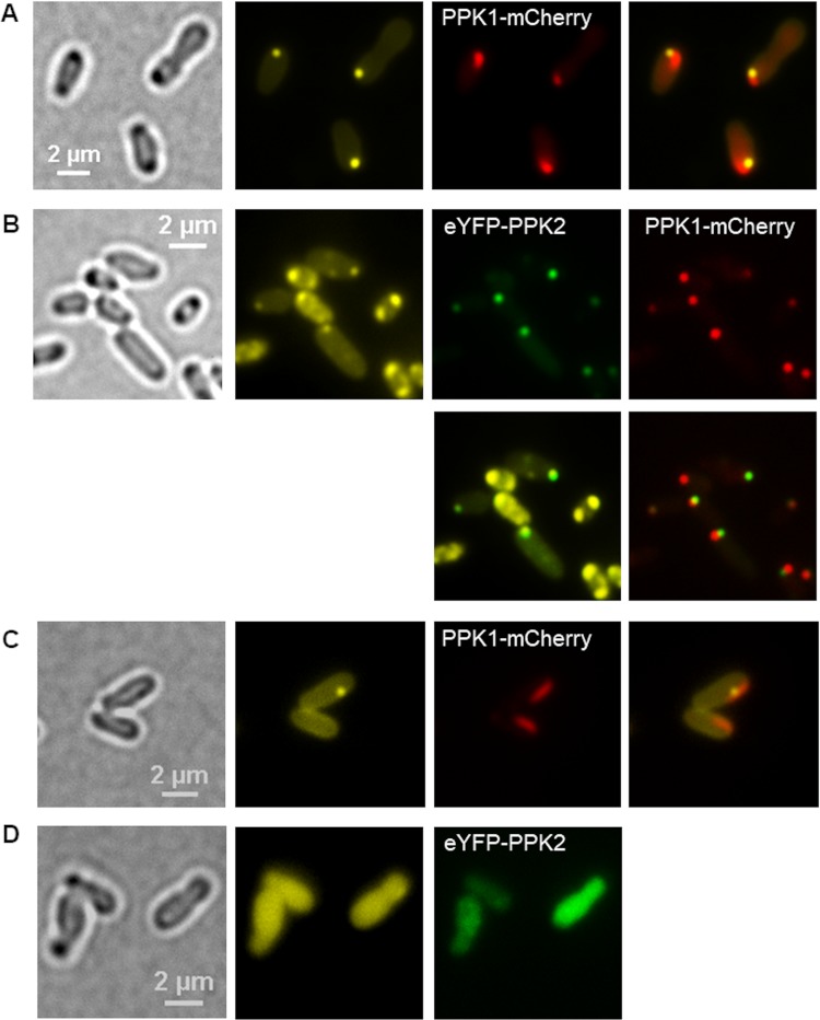
Localization of PPK1 and PPK2 in A. tumefaciens. (A) LB-grown cells of A. tumefaciens harboring pBBR1MCS2-PphaC-ppk1-mcherry were stained with DAPI and imaged (from left to right) in bright field, in the DAPI-polyP channel, and in the mCherry channel. The experiment was performed in five biological replicates. (B) A. tumefaciens cells with a genome-integrated ppk1-mcherry gene and harboring pBBR1MCS2-PphaC-eyfp-ppk2 are shown. (C and D) Cells of the A. tumefaciens Δppk1 Δppk2 mutant into which pBBR1MCS2-PphaC-ppk1-mcherry plasmid (C) or pBBR1MCS2-PphaC-eyfp-ppk2 (D) had been transferred. Note that ppk1-mcherry, but not eyfp-ppk2, was able to complement the formation of DAPI-polyP granules. (B to D) Samples were taken from three biological replicates.

