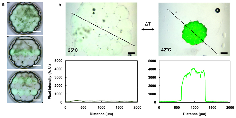
Figure 3. Patterned fluorescent PNIPAm hydrogel structures.
(a) Overlaid brightfield and fluorescence microscopy images of hexagonal hydrogel structures patterned with a fluorescent crosslinker, EBBA. Scale bars correspond to 500 μm. (b) Overlaid brightfield and fluorescence microscopy images, and pixel intensity plot profiles (along the black dashed lines) of fluorescent hexagonal hydrogel structures, before and after heating to 42°C. The pixel intensity increase is due to an increase in local EBBA concentration as a result of temperature-induced contraction. Scale bars correspond to 250 μm.

