
Figure 1.
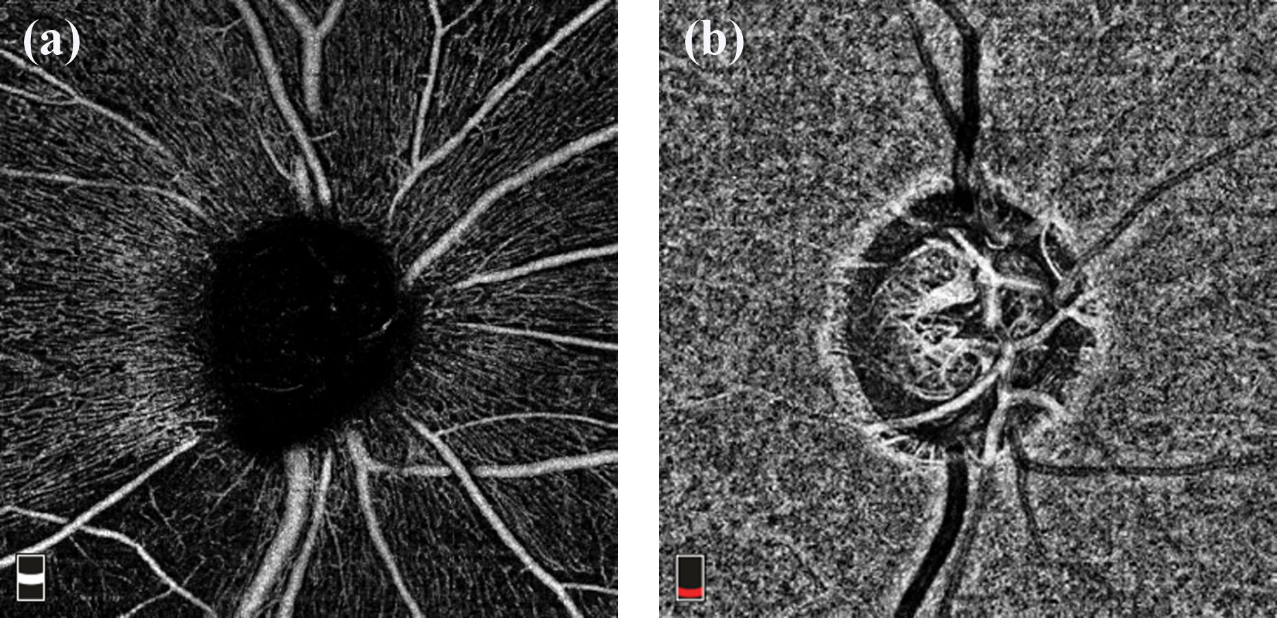
Angiography slabs of the optic nerve head scan obtained using spectral domain optical coherence tomography showing the radial peripapillary capillary, RPC (a) and choroid (b) slabs.
Official websites use .gov
A
.gov website belongs to an official
government organization in the United States.
Secure .gov websites use HTTPS
A lock (
) or https:// means you've safely
connected to the .gov website. Share sensitive
information only on official, secure websites.
Angiography slabs of the optic nerve head scan obtained using spectral domain optical coherence tomography showing the radial peripapillary capillary, RPC (a) and choroid (b) slabs.