Figure 4.

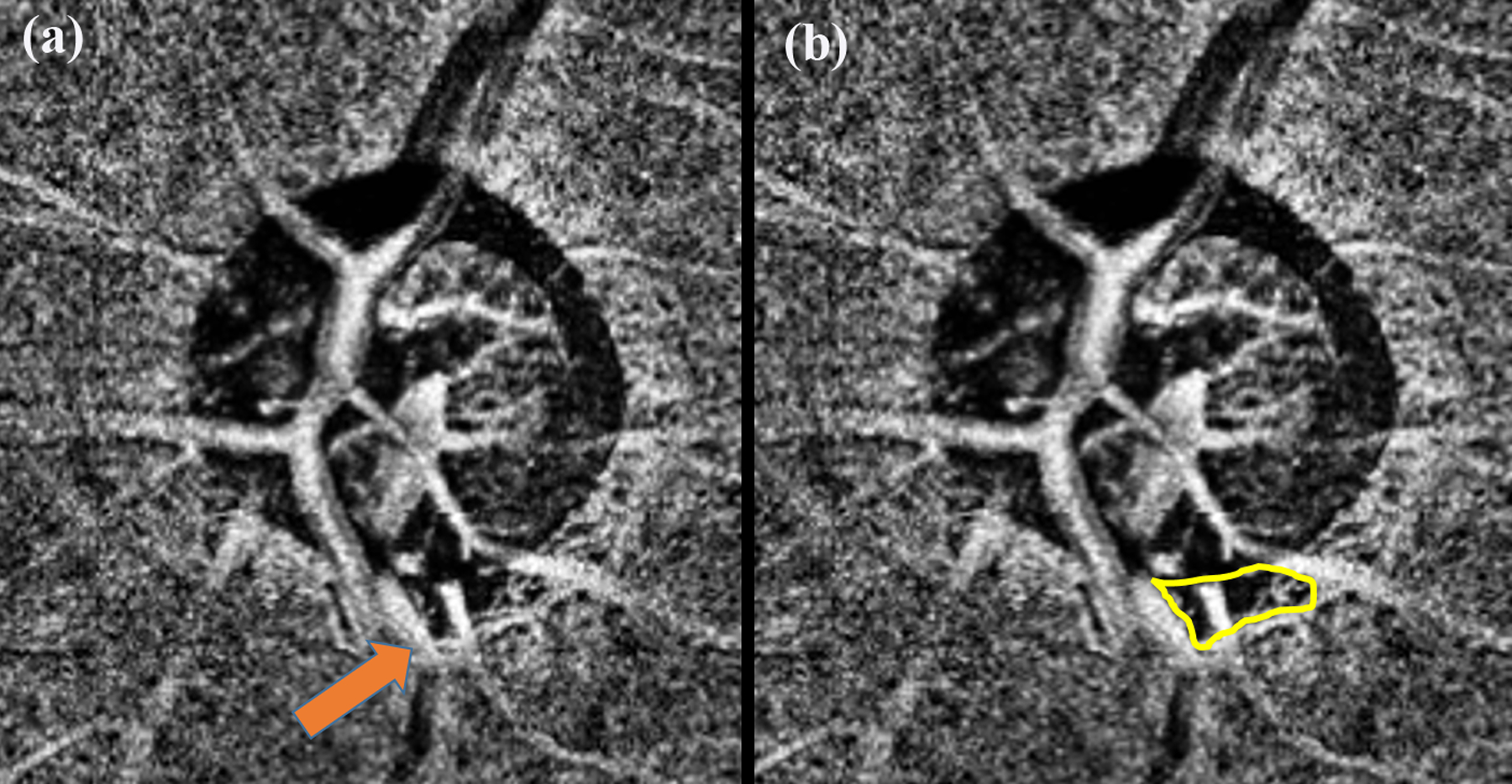
Choroidal OCTA slab of a glaucomatous eye showing the presence of deep-layer microvasculature dropout (MvD) in the inferior region (a). Arrow points to the MvD. Yellow line marks out the boundary of the MvD (b).

Choroidal OCTA slab of a glaucomatous eye showing the presence of deep-layer microvasculature dropout (MvD) in the inferior region (a). Arrow points to the MvD. Yellow line marks out the boundary of the MvD (b).