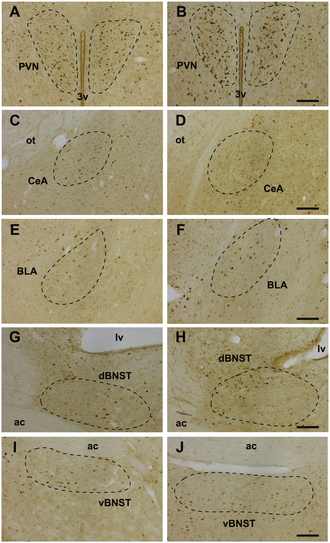
Fig. 2.

cFos immunoreactivity in various brain regions. The regions of interest are circled with dashed line. A, PVN of the cage-mate group. B, PVN of the pair-bonded group. C, CeA of the cage-mate group. D, CeA of the pair-bonded group. E, BLA of the cage-mate group. F, BLA of the pair-bonded group. G, dBNST of the cage-mate group. H, dBNST of the pair-bonded group. I, vBNST of the cage-mate group. J, vBNST of the pair-bonded group. 3v, the 3rd ventricle. lv, the lateral ventricle. ot, optic tract. ac, anterior commissure. Bars, 100 μm.
