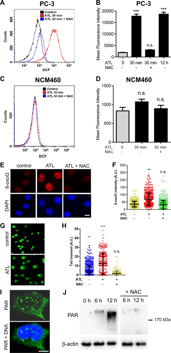
Fig. 1. A nontoxic dose of ATL induces oxidative DNA damage and activates PARP in cancer cells.
The PC-3 prostate cancer and NCM460 normal colon epithelial cells, with or without 1 h of preincubation in 10 mM NAC, were treated by 10 μM ATL or vehicle control for the indicated times. a, b Measurement of ROS in PC-3 cells by flow cytometry. Data from three independent experiments were presented as mean ± SD. c, d Measurement of ROS in NCM460 cells by flow cytometry. e, f Immunofluorescent staining of cellular 8-oxoG by Cy3-conjugated avidin. PC-3 cells were treated by 10 μM ATL for 12 h (scale bar: 10 μm). Nuclear 8-oxoG intensity was measured using the ImageJ software and the data were processed by the Prism software. g Representative images of alkaline comet assay. PC-3 cells were treated by vehicle control or 10 μM ATL for 12 h. h The tail moment was defined as percentage of tail DNA × tail length, quantified using the TriTek CometScore software. i Immunofluorescence staining for PAR foci in PC-3 cells treated by 10 μM ATL for 12 h. DNA was counterstained with DAPI (scale bar: 5 μm). j Western blot analysis of PAR in PC-3 cells. Ten micrometer ATL resulted in a time-dependent increase in PAR levels which was blocked by NAC. n.s. not significant, **p < 0.01, ***p < 0.001 vs. vehicle control.

