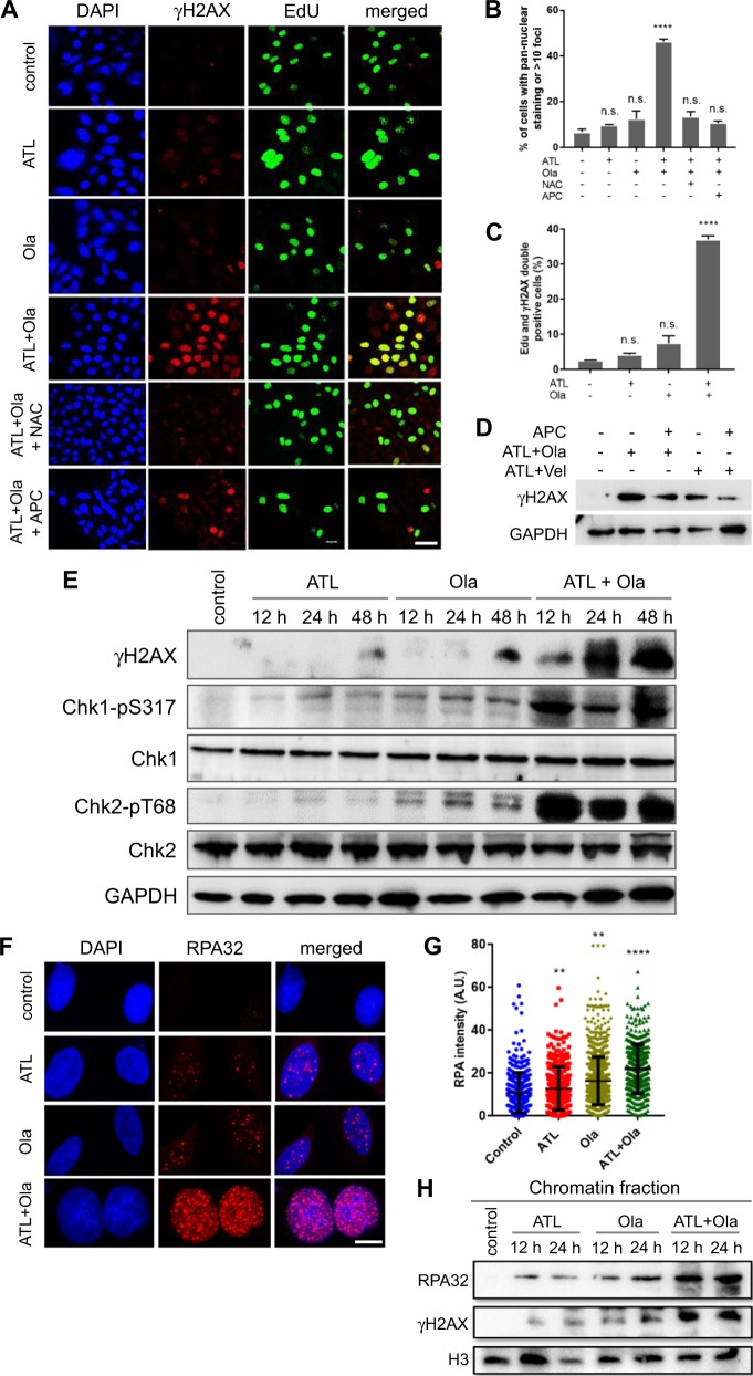
Fig. 4. ATL synergizes with olaparib to induce intense replication stress in cancer cells.
a Immunofluorescent staining of γH2AX and EdU. PC-3 cells were treated by 10 μM ATL, 10 μM olaparib (Ola) or the combination of the two, with or without 10 mM NAC or 5 μM aphidicolin (APC), for 12 h. At the end of drug treatment, cells were pulse-labeled with 10 µM EdU for 20 min (scale bar: 20 μm). b, c γH2AX and EdU positive cells were measured using the ImageJ software and the data were processed by the Prism software. d Western blot detection of γH2AX. PC-3 cells were treated by the combination of 10 μM ATL and 10 μM Ola or 10 μM ATL and 10 μM veliparib (Vel), with or without 5 μM aphidicolin (APC), for 24 h. e Western blot analysis of the indicated proteins. PC-3 cells were treated by 10 μM ATL, 10 μM olaparib (Ola) or the combination of the two for the indicated times. f Immunofluorescent staining of RPA32 foci. PC-3 cells were treated by 10 μM ATL, 10 μM olaparib (Ola) or the combination of the two for 12 h (scale bar: 10 μm). g Nuclear RPA32 intensity was measured using the ImageJ software and the data were processed by the Prism software. h Western blot detection of chromatin bound RPA32 and γH2AX. PC-3 cells were treated by 10 μM ATL, 10 μM olaparib (Ola) or the combination of the two for the indicated times. n.s. not significant, **p < 0.01, ****p < 0.0001 vs. vehicle control.

