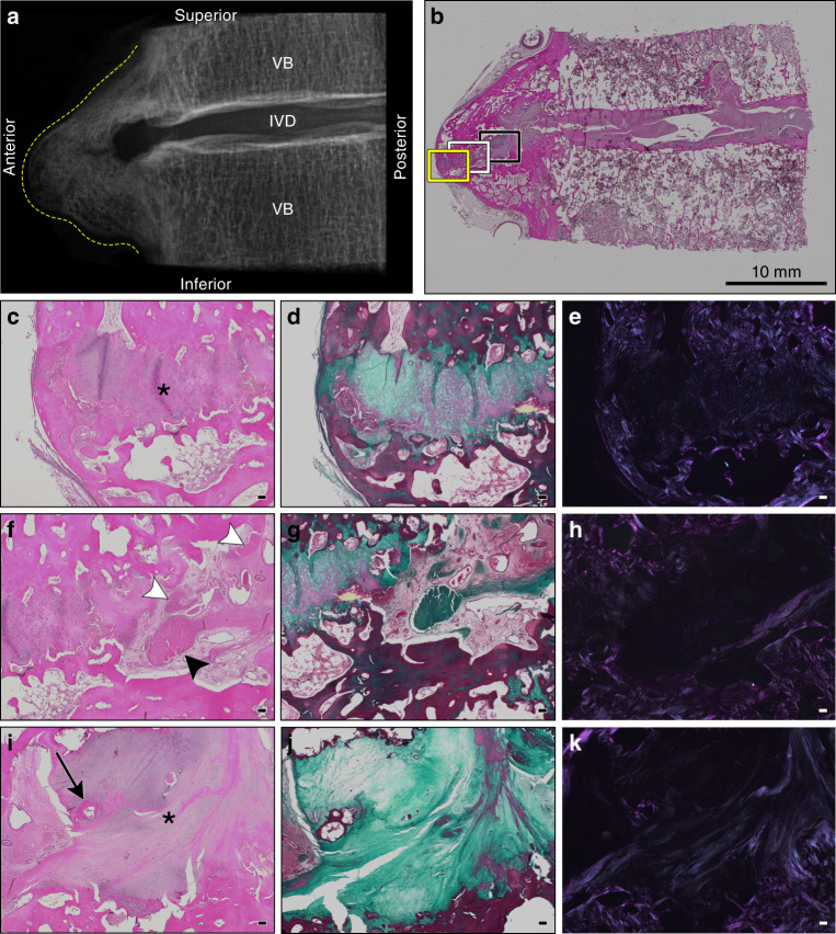
Fig. 3.
Histological appearance of a representative motion segment with horizontal and discontinuous-patchy presentations of ectopic mineralization associated with DISH. Images correspond to the specimen shown in Fig. 1c, T8-9. a Digital radiograph of the intact tissue prior to decalcification showing the localization of ectopic mineral within the motion segment (contoured by dotted yellow line). VB vertebral bone, IVD intervertebral disc. b Representative section stained with hematoxylin and eosin demonstrating the appearance of the intact section; scale bar represents 10 mm. Yellow (c–e), white (f–h), and black boxes (i–k) correspond to regions of fibrocartilage extending from the IVD positioned between mineralized outgrowths imaged with a 4× objective. c, f, i stained with hematoxylin and eosin; d, g, j stained with Masson’s trichrome; and e, h, and k the hematoxylin and eosin stained sections visualized with polarized light. Scale bars for c–k represent 100 µm. c–e Highlight the anterior-most portion of the fibrocartilaginous extension (asterisk). f–h Reveal a unique transition zone marked by the presence of amorphous granular material (black arrowhead) and multifocal areas of fibrosis (white arrowheads). i–k Display the fibrocartilage region (asterisk) adjacent to the native annulus fibrosus and a localized region of ossification (black arrow). All images are oriented as shown in a

