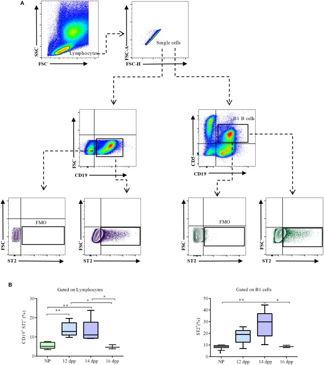
Figure 4.
Percentages of ST2-expressing total and B1 B cells in the peritoneal cavity of pregnant and non-pregnant mice. (A) Representative pseudocolor and contour plots showing gating strategy used to quantify percentages of ST2-expressing total CD19+ B cells as well as CD19+CD5+ B1 B cells in the peritoneal cavity of pregnant and non-pregnant mice. Lymphocytes were gated by FSC vs. SCC, doublets were eliminated, and CD19 alone or in combination with and CD5 were used to define total as well as B1 B cells, respectively. Fluorescence minus one (FMO) was used as control. (B) Box and whisker plots showing percentages of ST2-expressing CD19+ B cells in peritoneal cavity of non-pregnant (NP; n = 9) and pregnant mice at day 12 (12 dpp; n = 5), 14 (14 dpp; n = 9), and 16 (16 dpp; n = 3) of pregnancy as well as percentages of ST2-expressing CD19+CD5+ B1 B cells in non-pregnant (NP; n = 9) and in pregnant mice at day 12 (12 dpp; n = 5), 14 (14 dpp; n = 9), and 16 (16 dpp; n = 3) of pregnancy. Data are expressed as box and whisker plots showing median. *p < 0.05; **p < 0.01 as analyzed by ANOVA followed by Tukey's multiple comparisons test.

