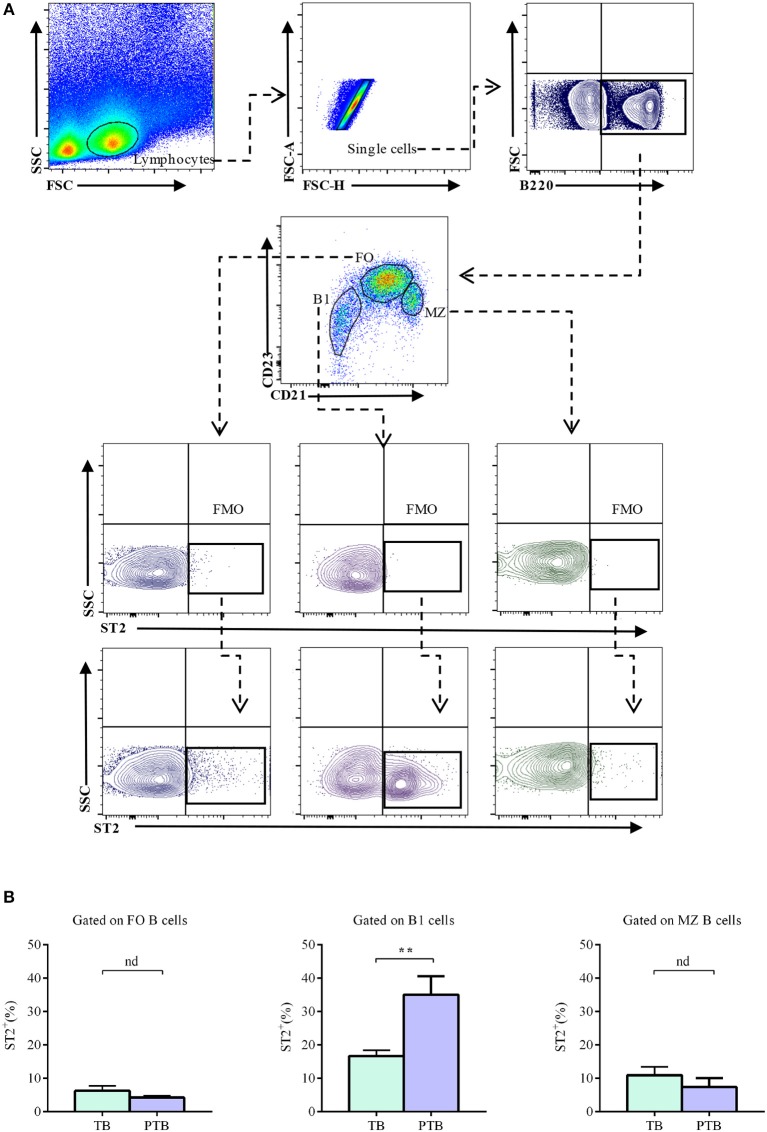
Figure 5.
Percentages of ST2-expressing follicular, marginal zone, and B1 B cells in the spleen of pregnant mice during the acute phase of LPS-induced preterm birth. (A) Representative pseudocolor and contour plots showing gating strategy used to quantify percentages of ST2-expressing follicular (FO), marginal zone (MZ), and B1 B cells in the acute phase of LPS-induced preterm birth and in term-pregnant control mice. Lymphocytes were gated by FSC vs. SCC, doublets were eliminated and B220 was used to define total B cells. CD23 and CD21 markers were used to identify FO (B220+CD23hiCD21low), MZ (B220+CD23lowCD21hi) and B1 (B220lowCD23−CD21−) B cells. Fluorescence minus one (FMO) was used as control. (B) Bar graphs showing percentages of ST2-expressing FO, MZ, and B1 B cells in preterm birth (PT; n = 6) and term birth control mice (TB; n = 6). Data are expressed as mean ± SEM. **p < 0.01 as analyzed by two-tailed unpaired t-test.

