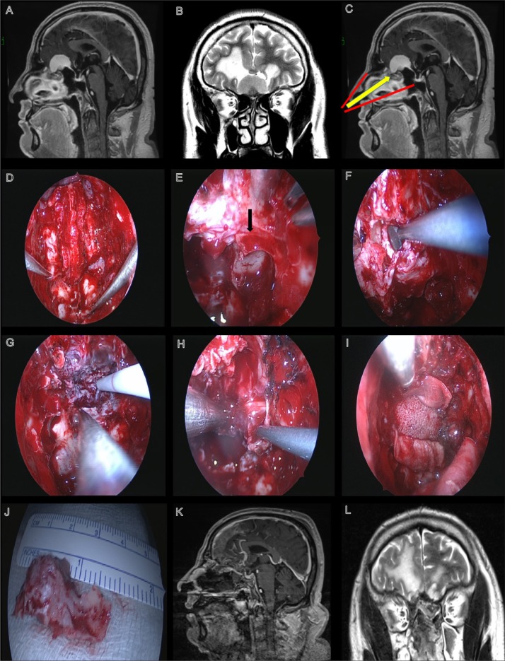
Figure 2.
Patient was a 50-year-old male presenting with seizures and anosmia. Preoperative T1-weighted image with gadolinium showing an olfactory groove meningioma that is homogeneously enhanced (A). Preoperative T1-weighted image showing an olfactory groove meningioma (B). The extent of the exposure for the transnasal approach in this case (C). View seen post complete bilateral sphenoethmoidectomy, septectomy, and an endoscopic modified Lothrop procedure (D). Posterior ethmoid artery seen on the left side (black arrow) (E). Dissection of mucosa along the skull base (F). Intraoperative views seen during (G) and after tumor resection (H). View after initial reconstruction using a MEDPOR porous plate and fascia lata (I). Resected portion of the crista galli (J). Postoperative images showing the complete removal of the lesion and the position of the nasoseptal flap (K, L).

