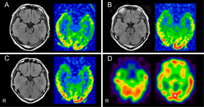
Figure 1.
Imaging findings in our patient and the characteristics of select cases of GABABR-AE. (A-C) FLAIR and ASL perfusion brain MRI sequences. Normal findings were obtained (A) at admission 12 days after syncope onset, at which point no neurological symptoms had been observed; (B) on day 14, at which point the patient had experienced repeated seizures; and (C) on day 38, after the patient’s limbic symptoms had markedly worsened. (D) Qualitative surface views of 123I-IMP perfusion SPECT images revealed no marked abnormalities on day 40. R: right, GABABR-AE: gamma-aminobutyric acid B receptor, FLAIR: fluid-attenuated inversion recovery, ASL: arterial spin labeling, 123I-IMP: 123I-N-isopropyl-p-iodoamphetamine

