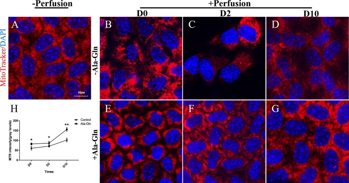
Figure 7.
Prevention of the decrease in the mitochondrial activity by Ala-Gln caused by intracameral irrigation. Representative images of MitoTracker (red) fluorescent staining in corneal endothelium pre-irrigation (A) and post-irrigation in the Ringer’s (B–D) or Ala-Gln (E–G) group at different time points.Nuclei were stained with DAPI (blue). (H) Relative fluorescence intensity of the mitochondria in corneal endothelium. Data are represented as mean ± SEM. *P < 0.05, **P < 0.01. (n = 3/group).

