
Fig. 8.
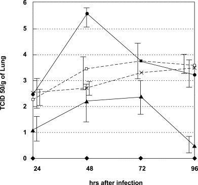
Virus titer in lungs of FluV A infected mice. Mice were infected in the same manner as described in the legend to Fig. 7. Three mice in each group were sacrificed at the indicated times, and their lungs were homogenized separately, diluted and centrifuged. Infective virus in the supernatant was titrated using MDCK cells and TCID50 was determined. ♦, noninfected control mice; ●, infected and untreated mice; □, mice treated with 2.4 mM PM-523; ×, mice treated with 40 mM ribavirin; and ▲, mice treated with 2.4 mM PM-523 plus 40 mM ribavirin.