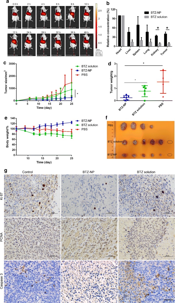
Fig. 4.
a The distribution of DiR labelled BTZ-NP in tumor bearing mouse measured by near-infrared fluorescence imaging (the black circle indicates the tumor tissue); b The ICP-MS results of the boron element distribution in the main organs and the tumors (* indicates statistically different, p < 0.05); c The tumor growth profiles (* indicates statistically different, p < 0.05); d The statistic results of the tumor weights at the end of the experiment (* indicates statistically different, p < 0.05); e The body weight curves; f The picture of the tumor tissues at the end of the experiment (the circle implies no tumor tissue observed); g IHC staining of the tumor tissues (the scale bar represents 50 µm)

