
Figure 2.
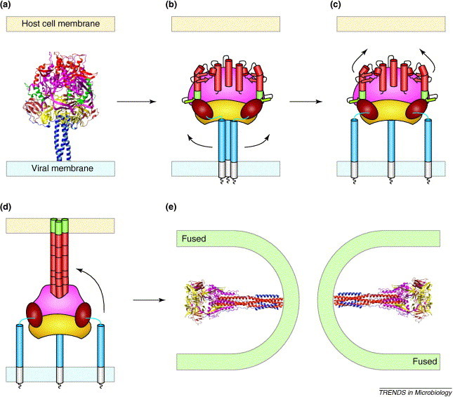
Model of the paramyxovirus-mediated membrane fusion. The F protein is thought to adopt five conformations during membrane fusion: (a) an inactive F0 precursor protein; (b) a metastable, native conformation of the cleaved F1+F2 protein; (c) an early fusion intermediate in which the HRB strands open up; (d) a pre-hairpin intermediate in which HRA forms a triple-stranded coiled coil and the fusion peptide inserts into the target membrane; and (e) the fusogenic hairpin structure that actively brings together the viral and cellular membranes. Ribbon diagrams are included of the pre-fusion PIV5 F0 protein structure [part (a), modified from [8]] and the post-fusion hPIV3 F protein structure [part (e), modified from [10]]. Conformations of the F protein that have not yet been determined at atomic resolution (b–d) are represented schematically as in Figure 1.