
Figure 2.
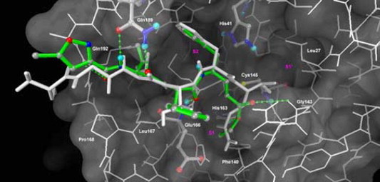
A modeled complex of SARS-CoV 3CL protease with the inhibitor 3c. The protease is shown in white line model, whereas the catalytic dyad (His41 and Cys145), His163 in S1 site, Glu166, Gln189 and compound 3c are highlighted in atom-colored ball-and-stick model. The stick of compound 3c is colored green. Individual atoms are displayed in gray (carbon), blue (nitrogen), red (oxygen), and yellow (sulfur). The hexapeptide Ser-Gly-Val-Thr-Phe-Gln (P6-P1), in white stick model, is overlaid for a comparison. The displayed structure of compound 3c is elected from the docked cluster with the lowest binding free energy. Potential hydrogen-bonding interactions of compound 3c with residues in the binding pocket are shown in green broken lines.