
Fig 2.
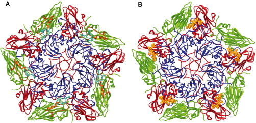
(A) Ribbon representation of a CS8c1 pentamer subunit (VP1, blue; VP2, green; VP3, red). The mobile antigenic G-H loop of VP1 (residues 130–160) is highlighted in yellow in a position corresponding to that found in the complex with the neutralizing antibody SD6 (Hewat et al., 1997) and in cyan for the position determined in the crystallographic structure of the reduced FMDV-O1BFS (Logan et al., 1993). The RGD integrin-binding triplet is depicted as sticks. (B) The structure of FMDV in complex with heparin (Fry et al., 1999). Heparin coordinates for five sugars are shown as yellow ball and sticks.