
Fig. 1.
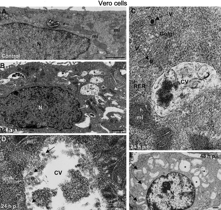
Associations of organelles in Vero cells infected with rubella virus. (A) Perinuclear areas in control, uninfected Vero cells, where mitochondria (mi) are randomly distributed. (B) Vero-infected cells at 16 h.p.i. exhibit cytopathic vacuoles (asterisks) surrounded by elongated, denser mitochondria (white arrows). (C) Higher magnification field showing a cytopathic vacuole (CV) attached to a mitochondrion (mi), rough endoplasmic reticulum (RER) cisternae, and a Golgi stack with viruses (V). (D) Cytopathic vacuole with vesicular structures containing dense spots (arrows). (E) Later in infection (48 h.p.i.) these associations of organelles mainly dissappear and in many cells both CVs (asterisks) and mitochondria (arrows) look empty. A, B, and D correspond to freeze-substituted cells, while C and E are conventionally processed samples. N, nucleus. Bars, 1 μm in A, B, and E; 200 nm in C and 100 nm in D.