
Fig 5.
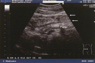
Transverse sonogram during an abdominal HAE attack, showing bowel and pancreas. Longitudinal section of a swollen bowel: the intestinal wall is edematously thickened (arrows); in addition, the reflectivity of the pancreas is increased.
Official websites use .gov
A
.gov website belongs to an official
government organization in the United States.
Secure .gov websites use HTTPS
A lock (
) or https:// means you've safely
connected to the .gov website. Share sensitive
information only on official, secure websites.
Transverse sonogram during an abdominal HAE attack, showing bowel and pancreas. Longitudinal section of a swollen bowel: the intestinal wall is edematously thickened (arrows); in addition, the reflectivity of the pancreas is increased.