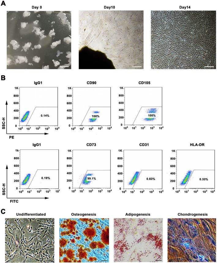
Fig. 1.
WJMSCs isolation and characterization. a Primary cell isolation procedure from Wharton’s jelly tissue. The migrated cells exhibited typical fibroblast-like morphology. Scale bar, 500 μm. b Flow cytometry analysis of P4 cells using mesenchymal stem cell markers (CD90, CD105, CD73), endothelial cell marker (CD31), and MHC class II protein HLA-DR. Isotypic antibodies (IgG1-PE and IgG1-FITC) were used as negative controls. c Representative stained images show that the fourth passage WJMSCs could differentiate into osteocytes (Alizarin Red S), adipocytes (Oil Red O), and chondrocytes (Alcian blue). Scale bar, 100 μm

