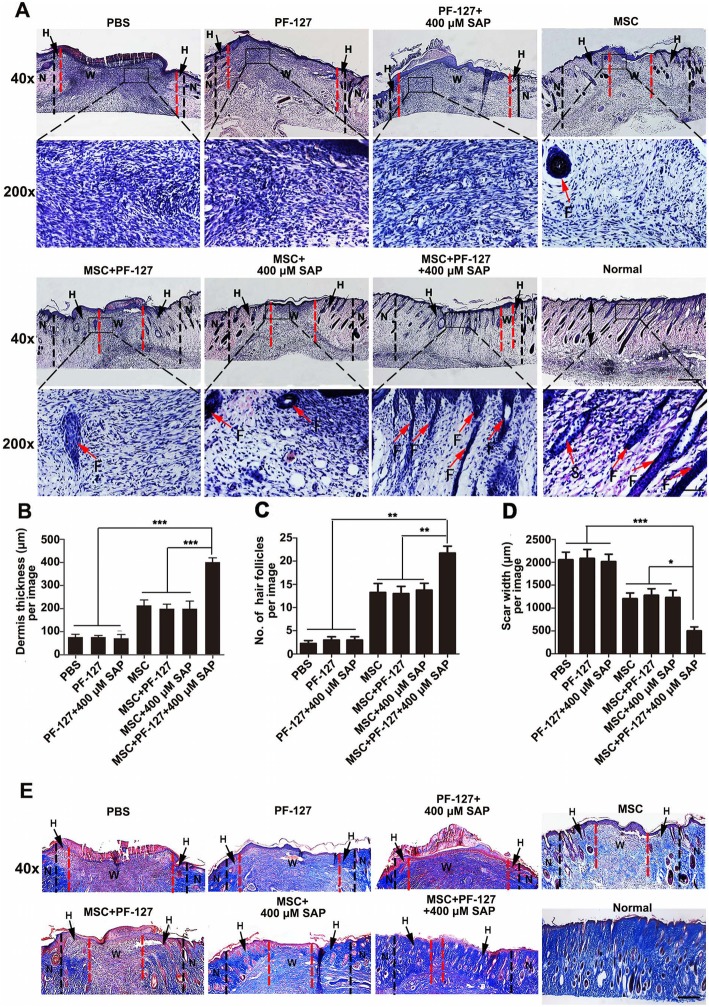
Fig. 4.
PF-127 plus SAP combination facilitates WJMSC-mediated dermis regeneration. a Hematoxylin-eosin (H&E) staining images of the wound bed and surrounding normal tissue (about 0.5 cm) at day 8 post-surgery. Scale bar: upper row, 500 μm; lower row, 100 μm. N, normal skin tissue, shown on both sides of the black imaginary line. H, healed skin tissue, shown between black and red imaginary lines. W, wound bed, unhealed skin tissue, shown between the red imaginary lines. F, hair follicle. b Quantitation data of healed dermis thickness at day 8 post-surgery. c Quantitation data of the number of newborn hair follicles at the healed site at day 8 post-surgery. d Quantitation data of scar width at the wound bed in different groups at day 8 post-surgery. In b, c, and d, data were presented as mean ± SD, n = 4. Statistical analyses were performed by one-way ANOVA followed by Tukey’s post-test. *p < 0.05, **p < 0.01, ***p < 0.001. e Masson’s trichrome staining images of the wound bed and surrounding normal tissue at day 8 post-surgery. Collagen fibers were stained dark blue. Scale bar: 500 μm. N, normal skin tissue, shown on both sides of the black imaginary line; H, healed skin tissue, shown between black and red imaginary lines; W, wound bed, unhealed skin tissue, shown between the red imaginary lines

