Figure 2.

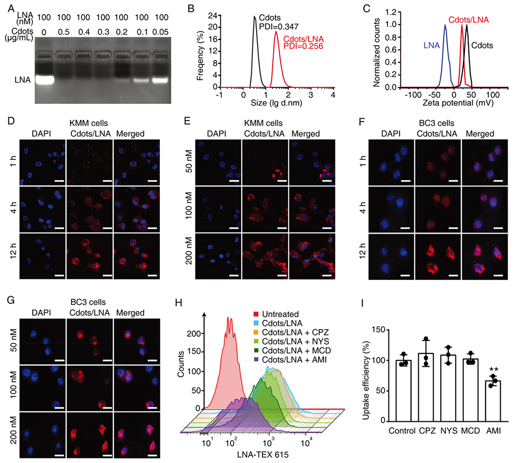
Efficient uptake of Cdots/LNA complexes by both adherent and suspension KSHV-infected cells. (A) Determination of the amount of Cdots required for loading 100 nM LNA analyzed by agarose gel electrophoresis. (B) Determination of the sizes of Cdots and Cdots/LNA complexes containing 1 μg/mL Cdots and 100 nM LNA by dynamic light scattering analysis (DLS). (C) Analysis of zeta-potentials of LNA, Cdots, and Cdots/LNA complexes. (D–G) Uptake of Cdots/LNA by KSHV-infected adherent KMM cells (D and E) and suspension BC3 cells (F and G) treated with Cdots/LNA for different times (D and F) and concentrations (D and G) analyzed by confocal laser scanning microscopy. LNA was conjugated with TEX615 dye, while the nuclei were stained with DAPI. Scale bars represent 20 μm. (H, I) Uptake efficiencies of Cdots/LNA by BC3 cells in the presence of different inhibitors including chlorpromazine hydrochloride (CPZ), nystatin (NYS), methyl-β-cyclodextrin (MCD), and amiloride (AMI) analyzed by flow cytometry (H) and the statistical analyses (I). LNA was conjugated with TEX615. Bars represent mean ± SD (n = 3). Statistical significance was calculated by one-way ANOVA with Tukey’s post hoc test. *P < 0.05; **P < 0.01; ***P < 0.001.
