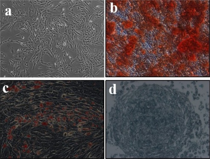
Fig. 1.
(a) The cell morphology of UC-MSCs was observed under a light microscope (magnification, ×100). (b∼d) Representative images of osteocyte, adipocyte, and chondrocyte differentiation of UC-MSCs cultured in the differentiation media. The cells were analyzed using cytochemical staining with Alizarin Red, Oil red O, and Alcian Blue respectively. The experiment shown is representative of three performed (magnification, ×200).

