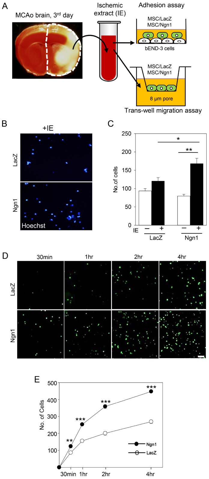
Fig. 2.
Enhanced adhesion and migration activity of mesenchymal stem cells expressing neurogenin 1 (MSCs/Ngn1). (A) Schematic diagram showing the experimental procedures of the adhesion and transwell migration assays. (B) Transwell migration assay showing Hoechst-stained mesenchymal stem cells expressing LacZ (MSCs/LacZ) and MSCs/Ngn1 in the bottom layer. (C) Quantification of Hoechst-stained cells that migrated to the bottom compartment containing the brain ischemic extract (IE) over a 4 h period. (D) Florescence image showing green fluorescent protein- (GFP-) positive MSCs/LacZ and MSCs/Ngn1 adhering to IE-stimulated bEnd.3 cells. (E) Quantification of GFP-positive cells following a 4 h adhesion assay. At least four random fields were used to obtain the number of adherent cells. Data are presented as mean±S.E. from three independent experiments. Statistically significant differences between MSCs/Ngn1 and MSCs/LacZ are indicated (*p<0.05, **p<0.01, ***p<0.001; Student’s t-test).

