
Fig. 5.
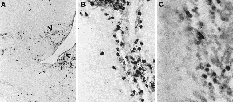
Cellular localization of NOS II protein in brains of acutely infected mice. Immunohistochemical staining of brain sections utilizing a polyclonal NOS II antibody followed by counterstaining with methyl green was performed on brain sections from mice sacrificed 3–7 days after infection. (A, B) NOS II positive cells in a brain 5 days after infection. Positive cells are seen surrounding blood vessels and migrating into the parenchyma. Vessels are indicated by arrowheads in (A). (C) F4/80 positive cells have a similar pattern of staining to that seen in B. Magnifications: (A) 25×, (B) 250×, (C) 400×.