
Fig. 7.
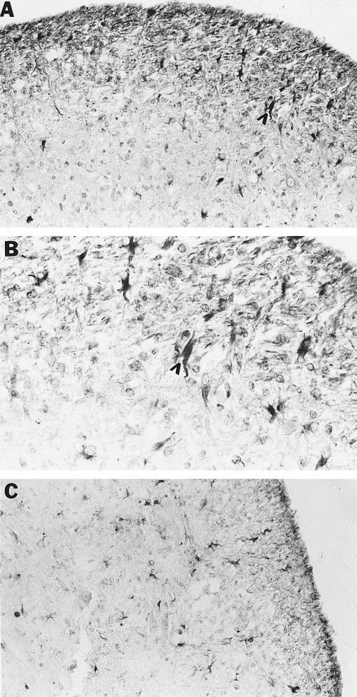
Cellular localization of NOS II protein in spinal cords of mice persistently infected with MHV-JHM. Immunohistochemical staining of brain sections utilizing a polyclonal NOS II antibody was performed on spinal cord sections of mice with hindlimb paralysis and chronic-active demyelinating lesions in the spinal cord. (A, B) NOS II positive cells were present in the lesion and around the lesion edge (B is a higher magnification of A). Arrowheads indicate one of many positive cells morphologically resembling astrocytes. (C) GFAP staining reveals a similar pattern to that seen in (A). Magnifications: (A, C) 100×, (B) 250×.