
Fig. 4.
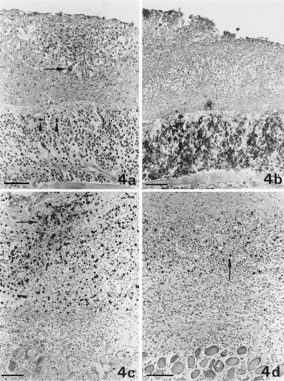
Diffuse alteration at the serosal surface of the diaphragm. (a) Expression of coronavirus antigen in single macrophages (granular reaction; arrow) within a granuloma with area of necrosis; single plasma-cells positive for coronavirus-specific antibodies (homogeneous cytoplasmic reaction; arrowheads), are seen in the underlying layers of lymphocytes and plasma-cells. Bar=60 μm. (b) Layers of precipitated exudate with a granuloma are separated from the unaltered parenchyma by a thick layer of CD45R antigen-positive B cells and plasma-cells. Bar=40 μm. (c) Granuloma comprised mainly of macrophages, expressing the myeloid/histiocyte antigen (arrow) within the layers of exudate. Bar=100 μm. (d) CD3 antigen-positive T-cells (arrow) are rarely seen scattered throughout the lesion. Bar=100 μm.