
Fig. 20.
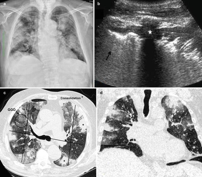
Integrated imaging (CXR, US, MDCT) in a nonsmoking, previously healthy 35-year-old man was presented in the emergency room after an unintentional exposure to chlorine gas at a community swimming pool. (a) CXR on presentation shows patchy bilateral lung consolidations. (b) Complementary LUS axial scan on the anterior chest at the fifth intercostal space shows focal consolidation (star) and aerated lung (arrow), without pleural effusion. MDCT (c) axial and (d) coronal reconstruction scans better demonstrate the extent of focal consolidations (*) and GGO areas; in (c) it has shown an example of radiologist’s score (RADS) findings in chest CT axial scan (see text)