
Fig. 18.
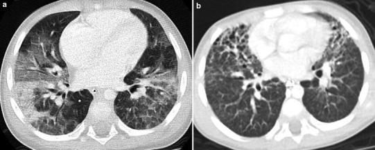
Acute interstitial pneumonia. An axial chest CT image (a) from a 2-year-old with fever and acute respiratory failure shows patchy bilateral ground-glass opacities and consolidation related to diffuse alveolar damage. An axial chest CT image (b) obtained 3 weeks later reveals traction bronchiectasis and architectural distortion of the anterior nondependent lung regions related to fibrosis