Fig. 3.

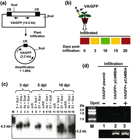
The Southern and PCR-based analysis to distinguish between the replicated and unreplicated forms of pCAMBIA/VA/GFP DNA. (a) The schematics of the experiment. Arrows represent the primers designed for the PCR and the indicated ScaI restriction sites are used for Southern analysis. (b) Diagrammatic representation of agro-infiltration of VA/GFP (upper panel) and time kinetics of GFP fluorescence in the infiltrated zone in tobacco leaf at 0, 3, 10, 15, and 20 d.p.i. (lower panel). Fluorescence was observed by using a low-magnification objective LWD 20C0.40 ph1 of an inverted fluorescent microscope (Nikon, Eclipse TE2000U). (c) Southern analysis of ScaI digested genomic DNA isolated from agro-inoculated tobacco. (d) Detection of circular amplicon by PCR. The plasmid DNA of the amplicon (Cam-VA/GFP), isolated from E. coli, was taken as a negative control for PCR using the primers mentioned in Section 2.4 (lane 1). The genomic DNA isolated from Cam/VA infiltrated leaf after 7 d.p.i., without (lane 2) and with DpnI digestion (lane 3), was taken as a template of the PCR for 21 cycles. The lower panel shows the respective amplification of actin as control.
