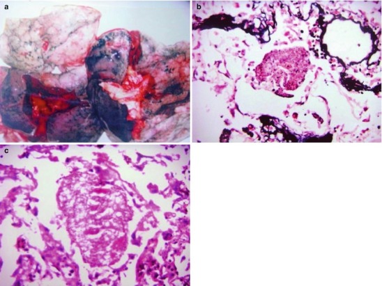
Fig. 17.1.

(a) Gross specimens’ observation demonstrates foamy liquid filling in the lung tissues. (b) HE demonstrates pneumocystis in the alveolar exudates, which can be stained black by silver methenamine staining, ×400. (c) HE demonstrates the foamy substance in the alveolar space, ×400
