Fig. 17.113.


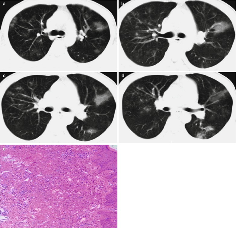
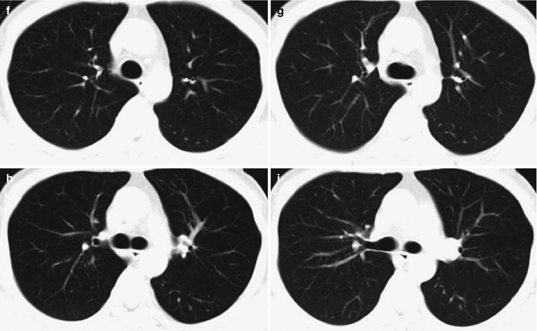
(a–e) HIV/AIDS related Kaposi’s sarcoma. (a–d) Chest CT scanning demonstrates scattered cloudy, mass and flake liked or nodular shadows with increased density. (e) Pathological biopsy demonstrates large quantity heteromorphological spindle cells with large thick stained nucleoli, which are in line with the diagnosis of Kaposi’s sarcoma. (f–i) Cured HIV/AIDS related Kaposi’s sarcoma. (f–i) Reexamination after treatment demonstrates absent lesions in both lungs, with clear lung fields
