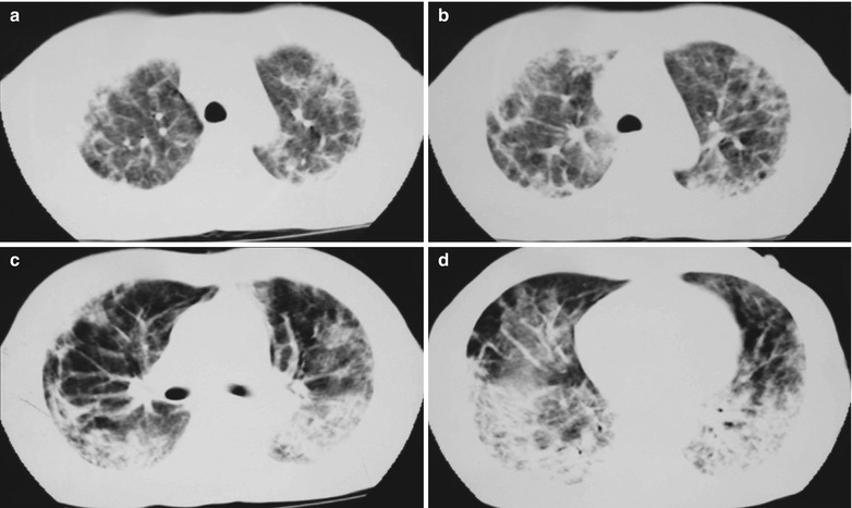
Fig. 17.12.

(a–d) HIV/AIDS related Pneumocystis carinii pneumonia. (a–d) CT scanning demonstrates multiple patchy parenchyma shadows and fibrous cords liked shadows in both lungs which are more obvious in the dorsal segment of both lower lungs, bronchial shadows in them, and thickened bronchial walls in the middle lobe. The hilar shadows in both lungs are enlarged
