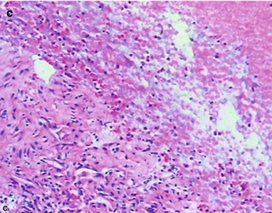
Fig. 17.36.


(a–c) HIV/AIDS related infiltrative pulmonary tuberculosis. (a) Pulmonary CT scanning of the pulmonary window demonstrates parenchymal shadows in the left lingual lobe with surrounding pulmonary acinar nodular shadows. (b) CT guided pucture biopsy of left lingula. (c) The pathology demonstrates granulation tissue and caseous necrosis, being in consistency with tuberculosis changes. HE × 100
