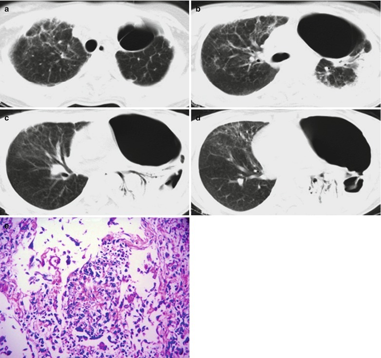
Fig.17.44.

(a–e) HIV/AIDS related pulmonary nontuberculous mycobacterial infection. (a–d) CT scanning demonstrates multiple cavities in the left lung field, bilateral multiple lobular central nodules and extensive branches liked linear shadows in tree buds sign. There are also large flaky parenchymal changes of the lung tissues in the left lower lung field in high density shadows, with accompanying air bronchogram sign. (e) HE staining demonstrates avium intracellular complex mycobacteria infection of lung tissues in atypical tuberculous nodular changes. (HE × 200)
