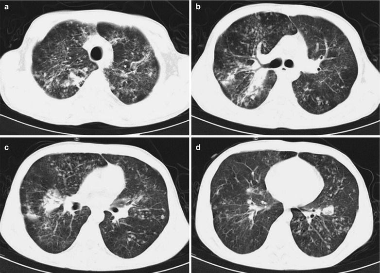
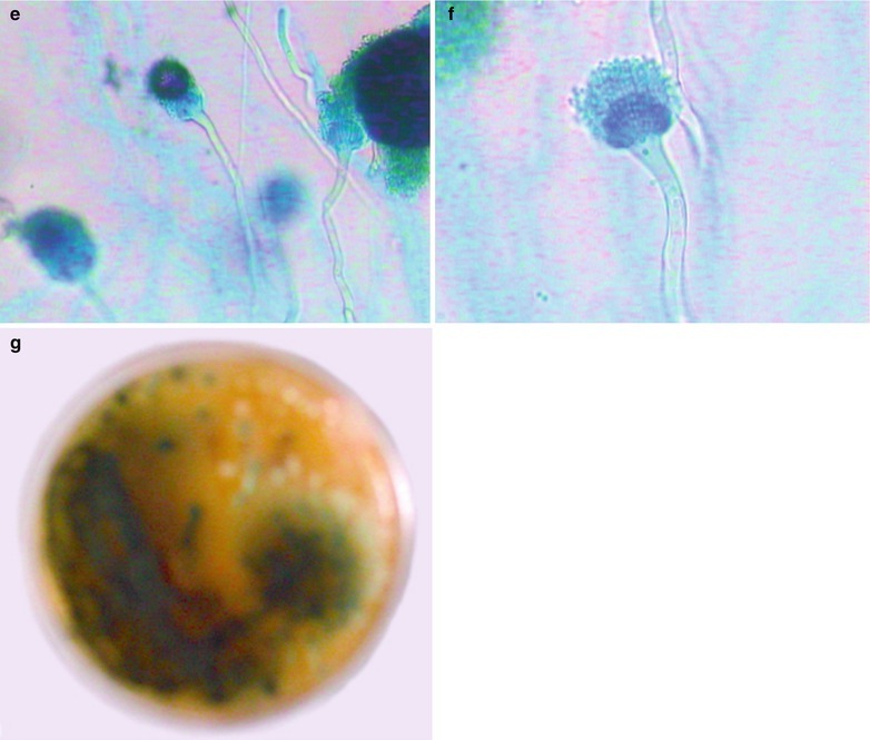
Fig. 17.76.


(a–g) HIV/AIDS related pulmonary aspergillosis infection. (a–d) CT scanning demonstrates diffuse scattered thin ground glass liked, patchy, flaky blurry shadows and cords liked shadows in both lungs, with blurry boundaries and uneven density; scattered nodular shadows in different sizes; more lesions in both upper lobes and the right middle lobe; flaky parenchyma shadows in the apical and posterior segments of right upper lobe, with air bronchogram sign in them; unobstructed opening of bronchi as well as lobar and segmental bronchi without stenosis and obstruction; lymphadenectasis in the right hilar region; detected Aspergillus fumigatus by sputum culture. (e, f) Culture for 72 h, lactic acid gossypol blue staining and microscopic observation at ×200 and ×400 demonstrate short column liked conidial head, smooth wall of conidiophores, flask-shaped top capsule and monolayer microconidiophores. (g) Culture in Paul’s medium demonstrates dark green colored colonies
