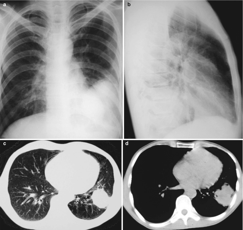
Fig. 17.80.

(a–d) HIV/AIDS related pulmonary cryptococcus infection. (a, b) Anteroposterior and lateral DR demonstrate a huge dense mass shadow in the left lower lung, with clear boundary. (c) CT scanning of the pulmonary window demonstrates round liked high density shadow in the left lower lung near left chest wall, with even density. (d) CT scanning of the mediastinal window demonstrates round liked soft tissue density shadows in the left lower lung near left chest wall, with even density, lobulation, and surrounding thick spikes
