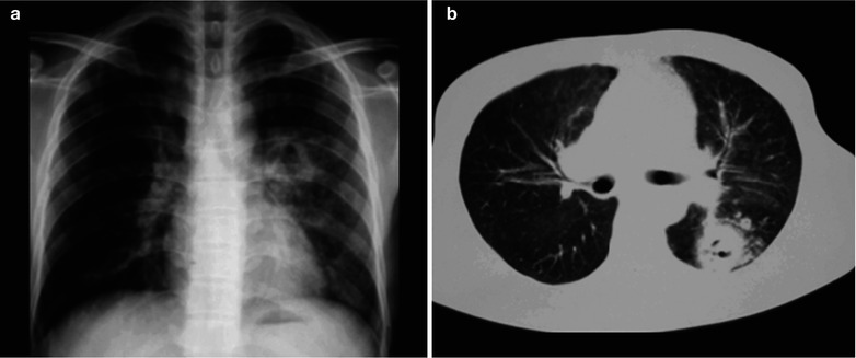
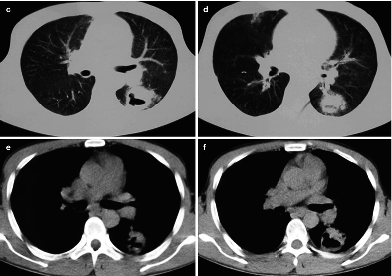
Fig. 17.85.


(a–f) HIV/AIDS related pulmonary cryptococcus infection. (a) DR demonstrates round liked thick-wall cavity in the left hilum, with blurred boundary; ground-glass liked shadows with increased density in the left middle and lower lung. (b–d) CT scanning of the pulmonary window demonstrates thick-wall cavity in the dorsal segment of the left lower lung, with uneven thickness of cavity wall. (e, f) CT scanning of the mediastinal window demonstrates thick-wall cavity in the dorsal segment of the left lower lung, with uneven thickness of the cavity wall and surrounding thick spikes
