Fig. 17.95.

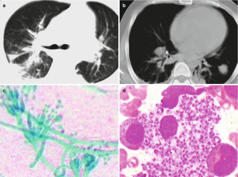
(a–d) HIV/AIDS related penicillium marneffei infection. (a) CT scanning of the pulmonary window demonstrates irregular large flaky shadows with increased density in the dorsal segment of both lower lobes; enlarged hilum in both lungs, cords liked thickening of the vascular vessels. (b) CT scanning of the mediastinal window demonstrates flaky parenchyma shadows in the left lower lung, thickening of both pleura, enlarged hilum shadows in both lungs, thickened right lower bronchial wall. (c) Microscopy after culture at 25 °C demonstrates branches and separated hyphae and its string of small spores, with typical penicillus but no sporangium (Medan staining, ×400). (d) Bone marrow smear demonstrates round or oval cells like the yeast phase within the macrophages; longer cells like the yeast phase outside the macrophages. The two kinds of cells have slightly curved ends in sausages liked appearance (HE staining, ×400)
