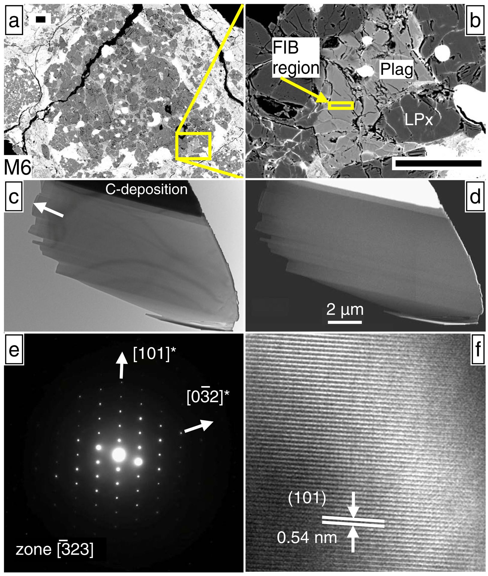
Fig. 6.

Electron micrographs of plagioclase from chondrule M6, which has resolvable excess 26Mg (Table 2; Fig. 4). (a) and (b) SEM images of plagioclase sectioned by FIB, where scale bars are 50 μm. (c) Bright-field TEM image of the plagioclase section. The sample appears as a single crystal, largely free of inclusions and defects, except for a small number of dislocations in the bottom right corner. The arrow corresponds to the surface of the meteorite thin section; about 1/3 of the FIB section was lost due to a pre-existing crack. (d) HAADF-STEM (Z-contrast image), showing the sample is chemically homogeneous (elemental EDS maps also show homogeneity in Electronic Appendix EA6). (e) Selected area electron diffraction (SAED) pattern consistent with only an unmodified anorthite structure and showing no superlattice reflections. (f) High resolution TEM (HRTEM) image, showing no inclusions are present, even down to the nm-scale.
