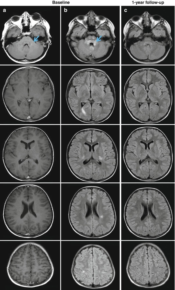
Fig. 5.2.
Brain axial post-contrast T1-weighted spin-echo (a) and fluid-attenuated inversion recovery (FLAIR) magnetic resonance images (b) from an 8-year-old patient with acute disseminated encephalomyelitis (ADEM). In (b), multiple hyperintense lesions involving both the white and gray matter are visible, whereas only one infratentorial lesion shows a mild enhancement (a, blue arrow). The one-year follow-up of the patient (c) shows a significant reduction of the size of hyperintense lesions on the FLAIR scan, without the formation of any new lesions

