
Fig. 1.
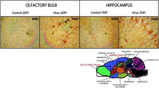
Illustration of the transneuronal route used by HCoV-OC43 for neuroinvasion and dissemination into the central nervous system. The left panel shows the olfactory bulb area, either mock-infected (control) or HCoV-OC43-infected (virus) at 3 days postinfection (DPI). The right panel shows the hippocampus, either mock-infected (control) or HCoV-OC43-infected (virus) at 7 days postinfection (DPI). In both regions of the brain, neurons are the target of infection. Magnification is 400X