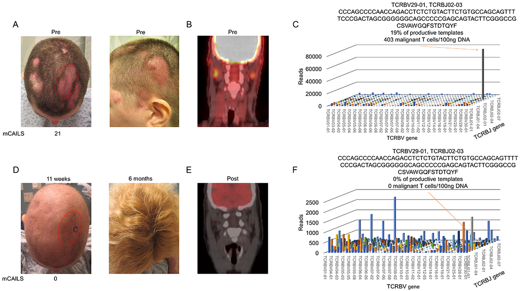
Figure 3. LDRT spares hair follicle destruction and can eradicate the malignant T cell clone in folliculotropic CTCL.

(A) Clinical photographs showing numerous erythematous plaques with overlying scale (left) with index lesion scored by mCAILS highlighted in red. Photo on right demonstrates thickness of plaques. (B) PET-CT scan showing PET-avid dermatopathic skin-draining lymph nodes prior to therapy. (C) 11 weeks after LDRT, clinical photograph showing anagen effluvium and clearance of original plaque stage disease (left) with 6-month follow-up picture (right) showing complete hair regrowth. (D) Repeat PET-CT scan after therapy showing resolution of PET-avid lymph nodes. (E) 3D histogram of TCRB sequencing showing V and J gene expression of hyperexpanded malignant T cell clone with resultant nucleotide and translated amino acid (AA) sequence of the CDR3 region. Based on productive templates, HTS revealed a malignant clone frequency of 19% and 403 malignant T cells/100ng DNA. (F) 3D histogram of TCRB sequencing shows eradication of the malignant T cell clone with recovery of benign T cell populations.
