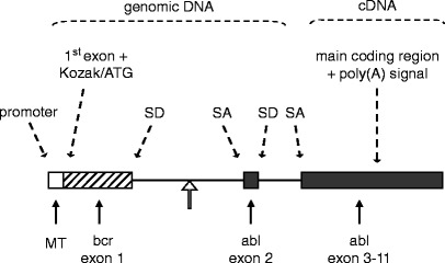
Fig. 3.

A textbook example of transgene design. The transgene depicted was constructed to study the development of Philadelphia-positive childhood leukemia in a mouse model. The Philadelphia chromosome, the hallmark of this clinical type of leukemia, results from a reciprocal translocation between chromosomes 9 and 22. As a result, the BCR locus and the ABL locus become joined. The genomic ABL locus itself is more than 200 kb in size, the first intron spanning approximately 175 kb. Breakpoints within the ABL locus are known to occur relatively 5′ within the first intron. The transgene harbors a heterologous (see Subheading 1.4) general-type metallothioneine promoter (24). The first BCR exon plus part of the first intron, which is fused (open arrow) to a short part of the first ABL introns and the second ABL exon, provide a splice donor (SD) and a splice acceptor (SA) site and preserve a simple but truthful mammalian intron–exon structure (see Subheading 1.2); an additional SD/SA pair comes from the ABL exon 2/intron 2 and intron 2/exon 3 boundaries. The main body of ABL exons, which spans about 32 kb, was cloned into the transgene as a cDNA segment. In this configuration, the transgene spans a mere 10 kb. Above illustration is adapted after (25).
