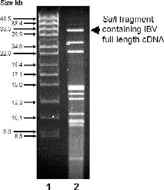
Fig. 5.

Analysis of SalI digested vaccinia virus DNA by PFGE. Lane 1 shows DNA markers and Lane 2 the digested vaccinia virus DNA. The IBV cDNA used does not contain a SalI restriction site; therefore the largest DNA fragment (∼31 kb) generated from the recombinant vaccinia virus DNA represents the IBV cDNA with some vaccinia virus-derived DNA at both ends.
